
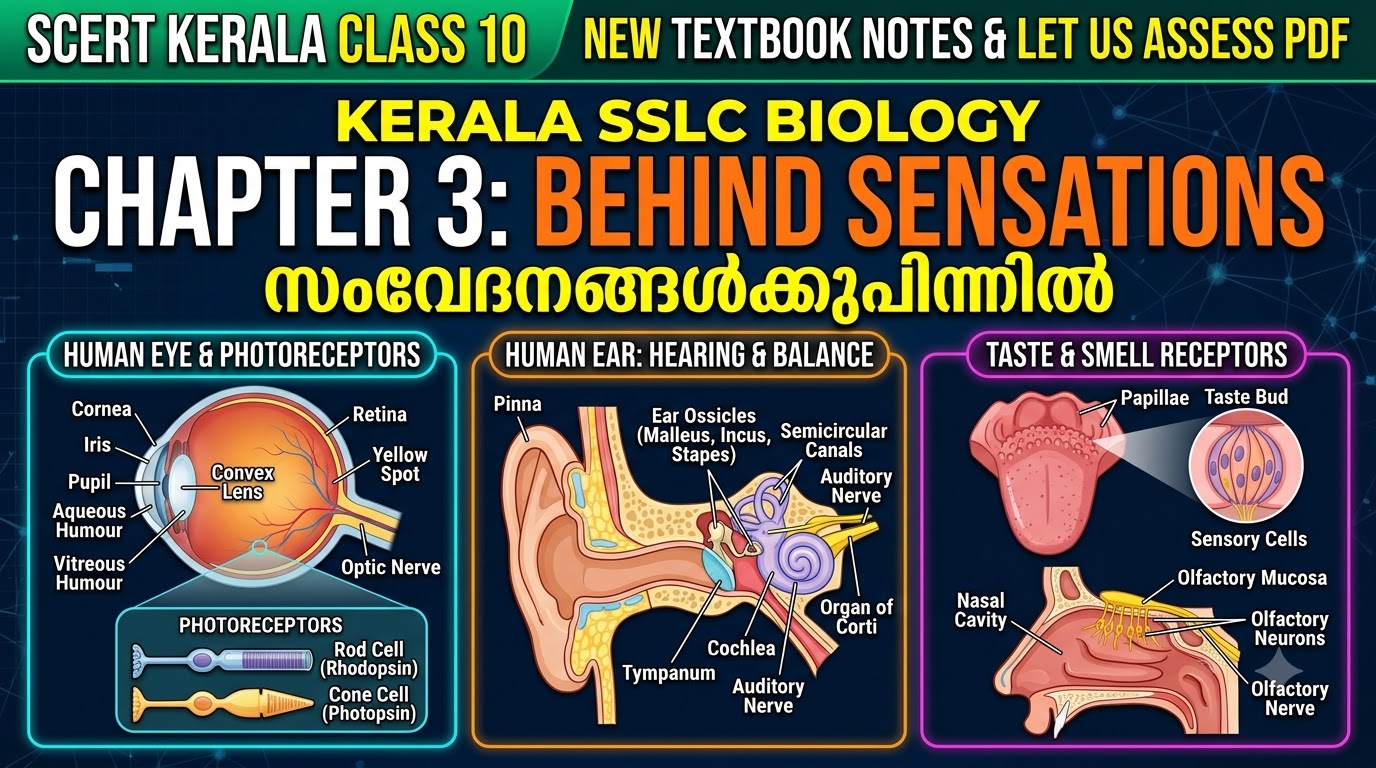
Kerala SSLC Biology Chapter 3 Notes for Behind Sensations (Malayalam Medium: സംവേദനങ്ങൾക്കുപിന്നിൽ) cover the third unit of the newly revised Class 10 (Std 10) Biology textbook published by the State Council of Educational Research and Training (SCERT Kerala). In the public examinations conducted by Kerala Pareeksha Bhavan, sense organs and the physiology of vision, hearing, equilibrium, taste, and smell consistently carry 5 to 7 marks, including compulsory flowchart and diagram-labelling questions on the Human Eye and Human Ear.
- 1. Structure of the Human Eye: 3 Layers, Chambers & Fluids (മനുഷ്യനേത്രത്തിന്റെ ഘടന)
- 1.1 Regulation of Pupil Size in Dim vs Bright Light (2-Mark Exam Question)
- 1.2 Comparison: Aqueous Humour vs Vitreous Humour & Power of Accommodation
- 2. Photoreceptors (Rod & Cone Cells), Chemistry of Vision & Eye Defects (പ്രകാശഗ്രാഹികളും നേത്രവൈകല്യങ്ങളും)
- 2.1 Major Eye Diseases & Defects (3-Mark Table)
- 3. Structure of the Human Ear: Mechanism of Hearing & Body Balance (ചെവിയുടെ ഘടനയും കേൾവിയും ശരീരതുലനനിലയും)
- 4. Taste, Smell, Skin Receptors & Sensory Organs in Other Organisms (രുചിയും ഗന്ധവും മറ്റ് ജീവികളിലെ സംവേദനാവയവങ്ങളും)
- 5. SCERT Textbook Let Us Assess Questions and Answers & Malayalam Medium Notes
As a High School Natural Science teacher, I have structured this complete English and Malayalam Medium guide to help you master every layer and chamber of the eye, the photoreceptors (Rod cells and Cone cells), the auditory pathway through the ear ossicles and cochlea, vestibular balance maintenance, and all solved Let Us Assess textbook questions.
Quick SSLC Biology & Science Navigation: Revise Chapter 1: Genetics of Life | Chapter 2: Paths of Evolution | Next: Chapter 4: Chemoreception in Organisms | SSLC Physics All 7 Chapters Hub | SSLC Chemistry All 7 Chapters Hub.
CHAPTER 3 AT A GLANCE: CLASS 10 BIOLOGY QUICK REVISION BLUEPRINT
- 3 Layers of Human Eye: Sclera (Outer white protective layer; transparent front = Cornea, covered by Conjunctiva), Choroid (Middle vascular layer with blood vessels; forms Iris & Pupil), Retina (Inner photoreceptor layer with Yellow Spot/Macula & Blind Spot).
- 2 Eye Fluids: Aqueous Humour (watery fluid between cornea & lens; formed from blood, nourishes cornea/lens) vs Vitreous Humour (jelly-like fluid between lens & retina; maintains eyeball shape).
- Photoreceptors: Rod Cells (pigment: Rhodopsin; dim-light B/W vision) vs Cone Cells (pigment: Photopsin/Iodopsin; bright-light & colour vision — Red, Green, Blue cones). Both pigments are synthesized from Vitamin A (Retinal + Opsin).
- Human Ear Dual Functions: Cochlea (Organ of Corti hair cells = Hearing) vs Vestibular Apparatus (3 Semicircular Canals + Vestibule = Body Equilibrium/Balance). Ear Ossicles: Malleus, Incus, Stapes (amplify vibrations ~22 times).
- Expected Board Weightage: 5 to 7 Marks across 1-Mark, 2-Mark, 3-Mark flowcharts, and 4-Mark diagram questions.
1. Structure of the Human Eye: 3 Layers, Chambers & Fluids (മനുഷ്യനേത്രത്തിന്റെ ഘടന)
Our eyes are protected inside deep bony sockets of the skull called Orbits and held in position by six external eye muscles. Tear secretion from the Lacrimal Glands contains the enzyme Lysozyme, which destroys germs and keeps the front of the eye moist.
| Layer / Part of Eye | Associated Structures | Exact Function (ധർമ്മം) |
|---|---|---|
| 1. Sclera (ദൃഢപടലം — Outer Layer) | Cornea (കോർണിയ) & Conjunctiva (നേത്രാവരണം) | White fibrous outer layer that gives firmness to the eye. The projected transparent anterior part is the Cornea, which refracts light rays into the interior of the eye. Conjunctiva covers and protects the front of the sclera except the cornea. |
| 2. Choroid (രക്തപടലം — Middle Layer) | Iris (ഐറിസ്), Pupil (കൃഷ്ണമണി), Ciliary Muscles & Convex Lens | Richly supplied with blood capillaries to provide oxygen and nutrients to the retina. The dark front part containing pigment Melanin is the Iris. The aperture at the centre of the iris is the Pupil. |
| 3. Retina (ദൃഷ്ടിപടലം — Inner Layer) | Yellow Spot / Macula (പീതബിന്ദു), Blind Spot (അന്ധബിന്ദു), Optic Nerve | Inner layer containing photoreceptors (Rod and Cone cells). Yellow Spot has maximum cone cells and is the point of keenest vision. Blind Spot is where the optic nerve begins; it has no photoreceptors and no vision. |
1.1 Regulation of Pupil Size in Dim vs Bright Light (2-Mark Exam Question)
- In Dim Light: The Radial Muscles of the iris contract and circular muscles relax, causing the Pupil to dilate (widen) so more light enters the eye.
- In Bright Light: The Circular Muscles of the iris contract and radial muscles relax, causing the Pupil to constrict (narrow) to protect the retina from intense light.

1.2 Comparison: Aqueous Humour vs Vitreous Humour & Power of Accommodation
| Feature | Aqueous Humour (അക്വസ് ദ്രവം) | Vitreous Humour (വിട്രിയസ് ദ്രവം) |
|---|---|---|
| Location | Filled in the Aqueous Chamber between the Cornea and the Lens. | Filled in the Vitreous Chamber between the Lens and the Retina. |
| Nature | Water-like fluid formed from blood and reabsorbed into blood. | Transparent jelly-like substance. |
| Function | Supplies oxygen and nutrients to the tissues of the Cornea and Lens (which lack blood vessels). | Helps maintain the spherical shape of the eyeball. |
Power of Accommodation (സമഞ്ജനക്ഷമത): The ability of the eye to form an image strictly on the retina by adjusting the curvature and focal length of the lens using Ciliary Muscles and Ligaments:
• While viewing Nearby Objects: Ciliary muscles contract → Ligaments relax → Curvature of lens increases → Focal length decreases.
• While viewing Distant Objects: Ciliary muscles relax → Ligaments stretch → Curvature of lens decreases → Focal length increases.
2. Photoreceptors (Rod & Cone Cells), Chemistry of Vision & Eye Defects (പ്രകാശഗ്രാഹികളും നേത്രവൈകല്യങ്ങളും)
| Feature | Rod Cells (റോഡ് കോശങ്ങൾ) | Cone Cells (കോൺ കോശങ്ങൾ) |
|---|---|---|
| Shape & Number | Rod-shaped; over 9 crore cells per retina. | Cone-shaped; around 45 lakh cells per retina (abundant in Yellow Spot). |
| Visual Pigment | Rhodopsin (Retinal + Opsin). | Photopsin / Iodopsin (Retinal + Opsin). Three types detect Red, Green, and Blue light. |
| Function | Activated in dim light; helps recognize objects in black and white (cannot distinguish colours). | Activated in bright light; provides colour vision and sharp details. |
| Related Disorder | Night Blindness (നിശാന്ധത) & Xerophthalmia (due to Vitamin A deficiency). | Colour Blindness (വർണ്ണാന്ധത) — hereditary defect of cone cells (cannot distinguish red & green). |
Chemistry & Flowchart of Vision (കാഴ്ചയുടെ രസതന്ത്രവും ഫ്ലോചാർട്ടും):
In the presence of light, visual pigments dissociate into Retinal (formed from Vitamin A) and Opsin (protein). This chemical change generates nerve impulses:
Light Rays → Cornea → Aqueous Humour → Pupil → Convex Lens → Vitreous Humour → Real, Inverted & Small Image on Retina → Rhodopsin/Photopsin dissociates into Retinal + Opsin → Photoreceptors generate Impulses → Optic Nerve → Visual Centre in Cerebrum → Experience of Erect 3D Vision (Binocular Vision)
2.1 Major Eye Diseases & Defects (3-Mark Table)
- Night Blindness (നിശാന്ധത): Inability to see clearly in dim light due to deficiency of Vitamin A, which prevents resynthesis of Retinal and Rhodopsin.
- Xerophthalmia (സീറോഫ്താൽമിയ): Prolonged deficiency of Vitamin A causes the conjunctiva and cornea to become dry and opaque, leading to permanent blindness.
- Colour Blindness (വർണ്ണാന്ധത): Genetic defect of cone cells causing inability to distinguish Red and Green colours; such persons are not selected as pilots, drivers, or railway signal operators.
- Glaucoma (ഗ്ലോക്കോമ): If the reabsorption of Aqueous Humour into the blood is obstructed, pressure inside the eye increases, damaging the retina and photoreceptor cells and causing permanent blindness (treated by laser surgery).
- Cataract (തിമിരം): The lens of the eye becomes opaque with age, obstructing light rays from reaching the retina (treated by replacing the defective lens with an artificial Intraocular Lens, IOL).
- Conjunctivitis (ചെങ്കണ്ണ്): Contagious infection of the conjunctiva caused by bacteria or viruses, spreading through touch and shared articles.
3. Structure of the Human Ear: Mechanism of Hearing & Body Balance (ചെവിയുടെ ഘടനയും കേൾവിയും ശരീരതുലനനിലയും)
| Region of Ear | Main Parts | Exact Function (ധർമ്മം) |
|---|---|---|
| 1. External Ear (ബാഹ്യകർണ്ണം) | Pinna (ചെവിക്കുട), Auditory Canal (കേൾവിക്കുഴൽ), Tympanum / Eardrum (കർണ്ണപടം) | Pinna directs sound waves into the auditory canal. Hairs and earwax trap dust and germs. Tympanum is a thin circular membrane that vibrates in resonance with sound waves. |
| 2. Middle Ear (മധ്യകർണ്ണം) | Ear Ossicles (Malleus, Incus, Stapes) & Eustachian Tube (യൂസ്റ്റേഷ്യൻ നാളി) | Ear ossicles amplify vibrations of the tympanum and transmit them to the Oval Window (Stapes is the smallest bone in the human body). Eustachian Tube connects the middle ear to the pharynx and balances air pressure on both sides of the tympanum. |
| 3. Internal Ear (ആന്തരകർണ്ണം) | Cochlea (കോക്ലിയ) & Vestibular Apparatus (3 Semicircular Canals + Vestibule) | Filled with fluids Perilymph and Endolymph. The Organ of Corti inside the snail-shaped Cochlea contains auditory hair cells for hearing. The Vestibular Apparatus contains hair cells for body balance. |
Flowchart of Mechanism of Hearing (കേൾവിയുടെ ഫ്ലോചാർട്ട് — 3 Marks):
Sound Waves → Pinna → Auditory Canal → Tympanum vibrates → Ear Ossicles (Malleus → Incus → Stapes) amplify vibrations → Oval Window → Vibration of Perilymph & Endolymph in Cochlea → Stimulation of Auditory Receptors (Hair Cells in Organ of Corti) → Impulses generated → Auditory Nerve → Cerebrum → Sense of Hearing
How the Ear Maintains Body Balance (ശരീരതുലനനില പാലിക്കുന്ന വിധം — 3 Marks):
Movement of the head causes movement of the Endolymph fluid inside the Vestibular Apparatus (Semicircular Canals and Vestibule: Utriculus & Sacculus). This stimulates the sensory hair cells inside the vestibular apparatus, generating nerve impulses that travel through the Vestibular Nerve to the Cerebellum, which coordinates muscular activities and maintains body equilibrium.
4. Taste, Smell, Skin Receptors & Sensory Organs in Other Organisms (രുചിയും ഗന്ധവും മറ്റ് ജീവികളിലെ സംവേദനാവയവങ്ങളും)
- Mechanism of Taste (രുചി അറിയുന്ന വിധം): Projected structures on the surface of the tongue are called Papillae, which contain Taste Buds (സ്വാദ് മുകുളങ്ങൾ) with chemoreceptors. Primary tastes: Sweet, Salt, Sour, Bitter, and Umami.
Flowchart:Food particles dissolve in Saliva → Stimulate Chemoreceptors in Taste Buds → Impulses generated → Nerve → Cerebrum → Experience of Taste - Mechanism of Smell (ഗന്ധം അറിയുന്ന വിധം):
Flowchart:Aromatic particles enter Nasal Cavity with inhaled air → Dissolve in Mucus → Stimulate Olfactory Receptors → Impulses generated → Olfactory Nerve → Cerebrum → Experience of Smell - Cutaneous Receptors in Skin (ത്വക്കിലെ ഗ്രാഹികൾ): Detect five sensations — Touch, Pressure, Pain, Heat, and Cold. Fingertips have the highest concentration of touch receptors.
- Sensory Receptors in Other Organisms (1-Mark Match-the-Following Table):
- Planaria & Euglena: Eye Spot (detects light).
- Housefly & Honeybee: Ommatidia (thousands of photoreceptors forming compound eyes).
- Snake: Jacobson’s Organ (vomeronasal organ on the roof of the mouth that detects aromatic particles brought in by the forked tongue).
- Shark & Fishes: Lateral Line (receptors along the sides of the body that detect water pressure variations and maintain balance).
5. SCERT Textbook Let Us Assess Questions and Answers & Malayalam Medium Notes
Q1. Why are we unable to see objects clearly for a few seconds when we suddenly enter a dimly lit cinema hall from bright sunlight?
Answer: In bright sunlight, the pupil is constricted to let in minimal light, and a major portion of the pigment Rhodopsin inside the rod cells is dissociated into Retinal and Opsin. When we suddenly enter a dark cinema hall, it takes a few seconds for the radial muscles of the iris to dilate the pupil and for Rhodopsin to be resynthesized from Retinal and Opsin so that rod cells can function in dim light.
Q2. Why do food items have very little taste when a person is suffering from a severe cold and blocked nose?
Answer: There is a close relationship between the perception of smell and taste. When a person has a cold, excess mucus blocks aromatic particles from reaching the olfactory receptors in the nasal cavity. Without the simultaneous sensation of smell, the brain cannot perceive the full flavour and taste of food.
Q3. During Eye Donation (Keratoplasty), is the entire eyeball of the donor transplanted into the recipient? Explain.
Answer: No. During eye donation, only the healthy transparent Cornea of the donor is surgically transplanted to replace the damaged/opaque cornea of the recipient (a procedure called Keratoplasty). The cornea should be collected within 6 hours of death.
പത്താം ക്ലാസ് ജീവശാസ്ത്രം അദ്ധ്യായം 3: സംവേദനങ്ങൾക്കുപിന്നിൽ (മലയാളം മീഡിയം നോട്ടുകൾ)
- കണ്ണിന്റെ 3 പാളികൾ: ദൃഢപടലം (Sclera) — മുൻവശത്തെ സുതാര്യമായ ഭാഗം കോർണിയ; രക്തപടലം (Choroid) — ഐറിസും കൃഷ്ണമണിയും ഉൾപ്പെടുന്നു; ദൃഷ്ടിപടലം (Retina) — പ്രകാശഗ്രാഹികൾ കാണപ്പെടുന്ന ആന്തരപാളി (ഏറ്റവും കൂടുതൽ കോൺ കോശങ്ങൾ ഉള്ള ഭാഗം: പീതബിന്ദു / Yellow Spot; പ്രകാശഗ്രാഹികൾ ഇല്ലാത്ത ഭാഗം: അന്ധബിന്ദു / Blind Spot).
- പ്രകാശഗ്രാഹികൾ: റോഡ് കോശങ്ങൾ (വർണ്ണവസ്തു: റൊഡോപ്സിൻ — മങ്ങിയ വെളിച്ചത്തിൽ കാഴ്ച നൽകുന്നു) vs കോൺ കോശങ്ങൾ (വർണ്ണവസ്തു: ഫോട്ടോപ്സിൻ — തീവ്രപ്രകാശത്തിലും വർണ്ണക്കാഴ്ചയും നൽകുന്നു). ജീവകം A-യിൽ നിന്നാണ് റെറ്റിനാൽ നിർമ്മിക്കപ്പെടുന്നത്.
- ചെവിയുടെ ധർമ്മങ്ങൾ: മധ്യകർണ്ണത്തിലെ യൂസ്റ്റേഷ്യൻ നാളി കർണ്ണപടത്തിന്റെ ഇരുവശത്തുമുള്ള വായുമർദ്ദം തുല്യമാക്കുന്നു. ആന്തരകർണ്ണത്തിലെ കോക്ലിയ (Organ of Corti) കേൾവിക്കും, വെസ്റ്റിബുലാർ അപ്പാരറ്റസ് (അർദ്ധവൃത്താകാര കുഴലുകൾ, വെസ്റ്റിബ്യൂൾ) ശരീരതുലനനില പാലിക്കുന്നതിനും സഹായിക്കുന്നു.
Teacher’s Classroom Note & Student Discussion Question:
Dear SSLC Students, always practice the two flowcharts in this chapter (Pathway of Light in Vision and Pathway of Sound in Hearing) — one of them is asked in almost every SSLC Biology Board Exam! Quick Question for You: Which tube connects the middle ear to the pharynx and balances air pressure on both sides of the tympanum? Write your answer in the comments below!


Latest SCERT & DHSE Kerala Study Notes
- Kerala Class 9 Physics Chapter 2 Notes: Equations of Motion (ചലനസമവാക്യങ്ങൾ) Std 9 Let Us Assess Questions and Answers PDF
- Kerala SSLC Christmas Exam Question Paper 2025-27: All Subjects Second Term & Model Question Papers with Solved Answer Keys PDF (New SCERT Syllabus)
- Kerala SSLC English Christmas Exam Question Paper 2025-27: Second Term & Model Solved 80-Mark Answer Key, Discourses & Grammar PDF
- Kerala SSLC Social Science Christmas Exam Question Paper 2025-27: Second Term & Model Solved 80-Mark Answer Key, Weightage & Map Study PDF
- Kerala SSLC Maths Christmas Exam Question Paper 2025-27: Second Term & Model Solved Answer Key, Weightage & Formula Sheet PDF (New SCERT Syllabus)
- Kerala SSLC Biology Christmas Exam Question Paper 2025-27: Second Term & Model Solved Answer Key, Weightage & Flowcharts PDF (New SCERT Syllabus)
- Kerala SSLC Chemistry Christmas Exam Question Paper 2025-27: Second Term & Model Solved Answer Key, Weightage & Sure A+ Questions PDF (New SCERT Syllabus)
- Kerala SSLC Physics Christmas Exam Question Paper 2025-27: Second Term & Model Solved Answer Key, Weightage & Formula Sheet PDF (New SCERT Syllabus)